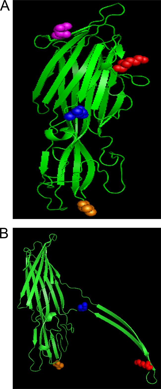
FIG 7.
Modeling CPB to the known structures of C. perfringens delta and NetB toxin. The secreted (A) and membrane-active (B) structure of CPB was predicted by modeling CPB to the resolved structure of C. perfringens delta and NetB toxin, respectively (29, 30). The CN3685 CPB amino acid sequence was threaded onto the model toxin using the PHYRE2 program. Once modeled, the locations of the 4 amino acid substitutions were identified and labeled with PyMOL software as follows: purple, K40N; red, E168K; blue, V191I; orange, A300V. Note that the K40N substitution was not modeled on Net B due to size differences between the toxins.

