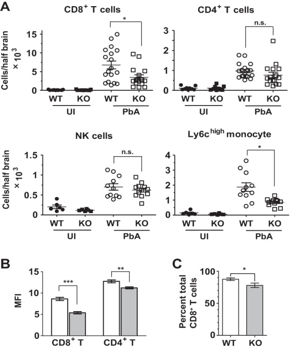
FIG 4.

Irgm3 deletion affects leukocyte recruitment to the brain. WT and Irgm3−/− (KO) mice were infected with 1 × 10⁶ PbA pRBC, and brains were collected following intracardiac PBS perfusion at day 6 or 7 p.i. (A) Flow-cytometric analysis of brain-sequestered leukocytes was performed. Symbols represent individual animals, and horizontal lines and error bars represent means ± SEM. Statistical analysis was performed via one-way ANOVA with Tukey's test. (B) Analysis of cell surface CD44 expression on brain-sequestered CD8+ and CD4+ T cells from PbA-infected WT and Irgm3−/− mice on day 6 or 7 p.i. Open bars represent the WT, and shaded bars represent Irgm3−/− mice. (C) Granzyme B intracellular staining in brain-sequestered CD8+ T cells from infected mice on day 7 p.i. The percentage of granzyme B+ cells is shown. Data presented are means ± SEM. Data are derived from two to three experiments with total n = 6 to 18/group (A and B) or a single experiment with 4 to 8/group (C). Statistical analysis was performed via unpaired t test (C) or two-way ANOVA with Bonferroni test (A and B). *, P < 0.05; **, P < 0.01; ***, P < 0.005; n.s, P > 0.05. UI, uninfected.
