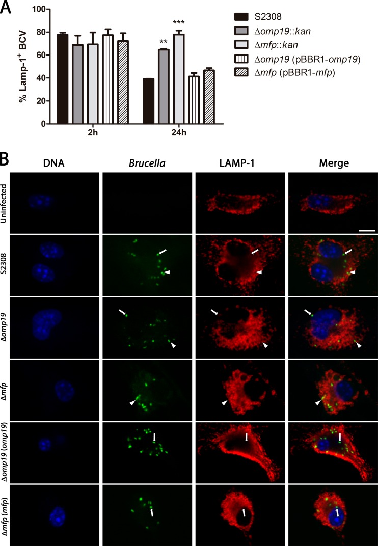
FIG 1.
Multiplication and intracellular localization of B. abortus Δmfp::kan, Δomp19::kan, Δmfp(pBBR1-mfp), Δomp19(pBBR1-omp19), and wild-type strains in BMDM. BMDM were infected (MOI of 100:1) with the B. abortus S2308(pBBR4-gfp), Δmfp(pBBR4-gfp), Δomp19(pBBR4-gfp), Δmfp(pBBR1-mfp, pBBR4-gfp), or Δomp19(pBBR1-omp19, pBBR4-gfp) strain. At 2 and 24 h postinfection, BCVs marked with LAMP-1 were directly enumerated by optical visualization using a confocal microscope. At least 50 BMDM were counted for each triplicate view. (A) Quantification of the percentage of wild-type or Brucella mutant BCVs containing LAMP-1 by confocal microscopy. Statistically significant differences in relation to the S2308(pBBR4-gfp) strain are indicated as follows: **, P < 0.01; ***, P < 0.001. (B) Representative confocal images of BMDM at 24 h postinfection with wild-type B. abortus or mutants. Brucella strains are labeled in green (GFP), LAMP-1 is shown in red (Alexa 546), and DNA is shown in blue (DAPI). Arrows point to Brucella bacteria outside LAMP-positive vesicles, and arrowheads indicate Brucella bacteria inside the LAMP-positive compartments. Scale bar, 10 μm.

