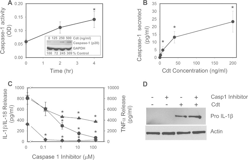
FIG 1.
Cdt-induced proinflammatory cytokine release from macrophages is dependent upon caspase-1 activation. (A) THP-1-derived macrophages were treated with 200 ng/ml Cdt for 2 and 4 h. Cell extracts were assessed for caspase-1 activity as described in Materials and Methods. The inset shows Western blot analysis of extracts obtained from THP-1-derived macrophages treated with 0 to 500 ng/ml Cdt; the mature form of caspase-1 (p20 subunit) is shown, along with glyceraldehyde-3-phosphate dehydrogenase (GAPDH) as a loading control. Levels of expression are shown below the blot as a percentage of levels in untreated control cells. OD, optical density. (B) THP-1-derived macrophages were treated with Cdt (0 to 200 ng/ml) for 5 h. Supernatants were then analyzed for the presence of caspase-1 (anti-p20) by an ELISA. Results are the means ± standard errors of the means for three experiments, each performed in triplicate. (C) Effects of caspase-1 inhibition on release of proinflammatory cytokines. Macrophages were exposed to various amounts of the caspase inhibitor Z-WEHD-FMK for 60 min; 50 ng/ml Cdt was then added to the cells for 5 h. Culture supernatants were analyzed for IL-1β (circles), IL-18 (diamonds), and TNF-α (triangles) by an ELISA. Results are plotted as the mean ± the standard error of the mean pg/ml cytokine released versus the Z-WEHD-FMK concentration. Levels of cytokines release from untreated control cells were 9.8 ± 7.8 pg/ml (IL-1β), 38.1 ± 14.0 pg/ml (IL-18), and 120.9 ± 2.6 pg/ml (TNF-α). Asterisks indicate statistical significance (P ≤ 0.05) compared to untreated control cells (A and B) or compared to cells treated with toxin alone (C). (D) Cells were assessed for the expression of pro-IL-1β after 5 h of exposure to Cdt following solubilization, fractionation by SDS-PAGE, and analysis by Western blotting. Actin is shown as a loading control. Western blots are representative of results from three experiments.

