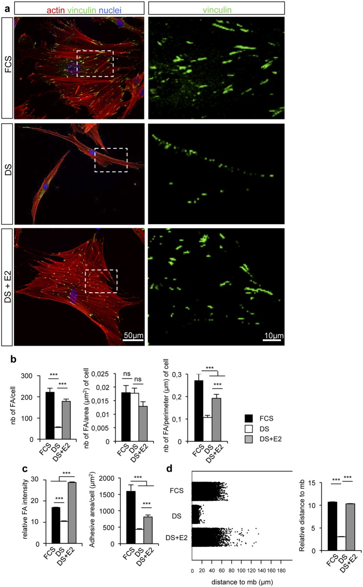
Fig 2. 17β-estradiol treatment correlates with focal adhesion strengthening.
a. Confocal pictures of donor 1 cultured in the presence of FCS, or DS supplemented or not with 10-7 M E2. Vinculin (green), actin (red) and nuclei (blue) were stained. b. Quantification of focal adhesions number, related or not to cell area or cell perimeter. c. Quantification of vinculin staining intensity and adhesive area by cell. d. Diagram of jitter plot (left) and mean (right) describing the distance of vinculin staining to the periphery of the membrane. Diagrams (except jitter plot) represent the mean±SEM of n>50 cells per condition. ***: p<0.001, ns: not significant.

