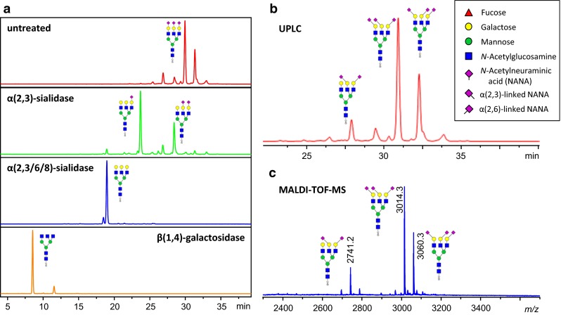
Fig. 1.

2-AB-labeled A3 glycan standard from Ludger (CAB-A3-01) was analyzed by UPLC and MALDI–TOF–MS after ethyl esterification. a Overnight incubation at 37 °C of Ludger 2-AB-labeled A3 standard with buffer (red), α(2,3)-sialidase (green), α(2,3/6/8)-sialidase (blue), α(2,3/6/8)-sialidase + β(1,4)-galactosidase (orange). Separation was performed on a Waters Acquity UPLC H-class system using an Acquity BEH 1.7 µm 2.1 × 150 mm glycan column. b Linkage-specific assignment of the undigested UPLC data [100]. c MALDI–TOF–MS spectrum of the 2-AB-labeled A3 standard ([M + Na]+) after 1 h of ethyl esterification with EDC and HOBt at 37 °C and subsequent HILIC purification according to Reiding et al. [43]. Profiles obtained from both methods are highly comparable and show similar ratios with regard to sialic acid occupancy and linkage. HILIC peak assignments are based on the exoglycosidase digests, as well as use of internal standards. Structural schemes of glycans are depicted following the CFG notation: N-acetylglucosamine (blue square), fucose (red triangle), mannose (green circle), galactose (yellow circle), N-acetylneuraminic acid (purple diamond). Known N-acetylneuraminic acid linkages are indicated by a left angle (α2,3) or right angle (α2,6), and otherwise unspecified
