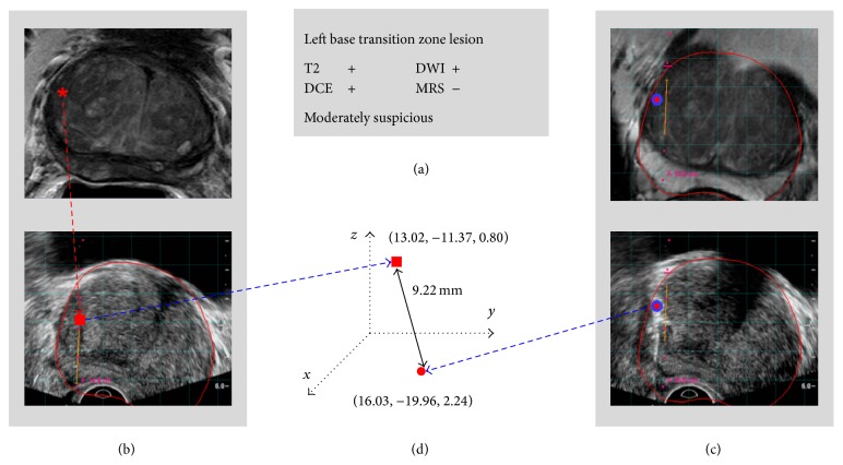
Figure 1.
Method for calculating target location difference between visual registration and semiautomated MRI-US fusion. (a) MRI detects lesions (MRI reading). (b) A suspicious lesion is visually correlated and estimated on the US image (red rectangle). (c) The lesion is also registered through MRI-US fusion system (red circle). (d) The spatial difference between the estimated target using just ultrasound and actual target using EM-based MRI-US fusion is calculated. T2: T2-weighted, DWI: diffusion-weighted, DCE: dynamic contrast-enhanced, and MRS: MR spectroscopy.

