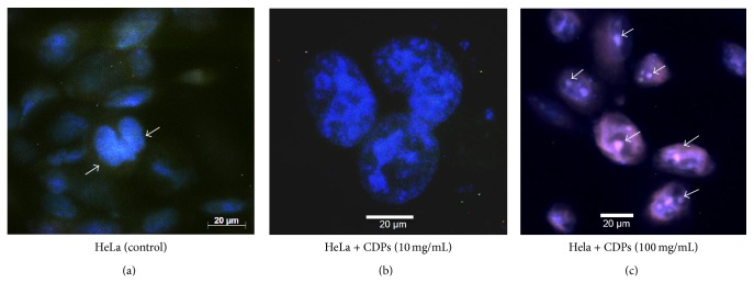
Figure 3.

Morphological changes in HeLa cells, induced by cyclodipeptides from Pseudomonas aeruginosa. ((a), (b)) Images of HeLa cells were taken under phase-contrast confocal microscopy following treatment with the P. aeruginosa CDP mix for 24 h and staining with DAPI. Images of cells were taken at 20x magnification (a) or 40x magnification (b). (c) Assessment of nuclear condensation by DAPI staining of cells treated with P. aeruginosa PAO1 CDPs (20x magnification). After treatment, the number of apoptotic nuclei was increased and more nuclear condensation was observed in cultures of both cell lines, in comparison with untreated controls. Arrows indicate apoptotic nuclei.
