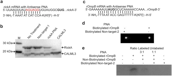
Fig 1. PNA is specific for designed target.
A) rickA PNA target with putative Shine Dalgarno region in red and start codon in bold B) Western blot of in vitro translation assay supplemented with PNA complementary to the cloned region of rickA, no PNA, or non-targeting control-1 PNA. In vitro translation reactions contained vectors coding both truncated RickA and the control CALML3, or CALML3 alone. C) rOmpB PNA target with start codon in bold D) Dot blot confirming biotinylation of rOmpB and non-targeting-2 PNA. E) Nylon membrane probed to detect biotinylated PNA-ssRNA pairs following rOmpB PNA target RNA denaturation and hybridization to biotinylated PNA, incubated with increasing ratios of unlabeled PNA for competitive binding assay. Incubations with either no PNA or biotinylated non-targeting-2 PNA serve as a control.

