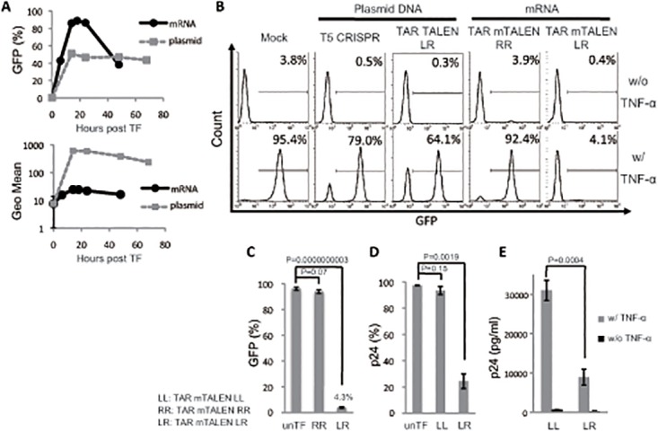
Fig 2. HIV LTR editing with mRNAs of TAR TALENs.

(A) Kinetics of GFP transduction with mRNA and plasmid DNA in Jurkat cells. Jurkat cells were transfected with 1 μg of mRNA GFP or 1 μg of plasmid GFP under the control of a CMV promoter. The time course analysis of GFP expression was performed by flow cytometry. (B and C) A Jurkat cell line latently transduced with an LTIG vector was transfected with mTALENs. The level of GFP expression 48 hours after TNF-α stimulation is shown. Representative histograms are shown in B. The positive percentage of GFP is shown in B (n = 3). (D and E) ACH-2 cells were transfected with TAR mTALENs. The expression of p24 antigen 48 hours after TNF-α stimulation is shown. The percentage of p24 antigen expression in cells is shown in C (n = 3). The amount of p24 antigen in the culture supernatant is shown in E (n = 3). The error bars in A, C, D, and E show standard deviations (n = 3).
