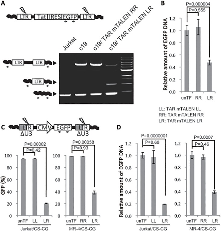
Fig 3. Excision of HIV provirus from host cell genome with TAR mTALENs.

(A and B) HIV proviral excision in c19. (A) HIV provirus in c19 treated with TAR mTALENs was amplified using a primer set designed for the host cell genome sequence flanking the proviral integration site. The schematic of PCR products indicating genomic sequences, full-length provirus, and one LTR footprint resulting from proviral excision are shown on the left side. (B) The relative amount of EGFP DNA in TAR mTALENs—treated c19 is shown. (C and D) Excision of HIV-based lentiviral vector DNA with TAR mTALENs. Jurkat and MT-4 cells were transduced with a lentivirus vector containing a CMV promoter—derived GFP-expressing cassette, Jurkat/CS-CG and MT-4/CS-CG cells, respectively, and, these cells were treated with TAR mTALENs. A schematic of the lentiviral vector DNA used in this assay is shown at the top of C. The percentage of GFP positive cells after TF of mTALENs is shown in C (n = 3). The relative amount of EGFP DNA in treated TAR mTALENs is shown in D (n = 3). The error bars in B, C, and D show standard deviations (n = 3).
