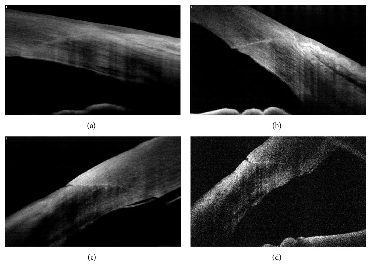
Figure 5.
Architectural analysis of cataract surgery. Well apposed corneal wound (1 plane) (a), loss of cooptation with minimal endothelial misalignment (2 plane) (b), minimal Descemet's membrane detachment with epithelial gap (2 plane) (c), and loss of cooptation with endothelial gap (3 plane) (d).

