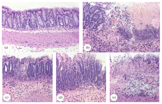
Figure 2.
Representative light microscopy of colons from both colitic and noncolitic groups. Micrographs of the groups saline (a), TNBS (b), and RJ 100, RJ 150, and RJ 200 receiving both TNBS and royal jelly (c), (d), and (e), respectively. Hematoxylin and Floxin staining. Micrographs were taken using 10x objective lens. Each image is representative of 3 animals.

