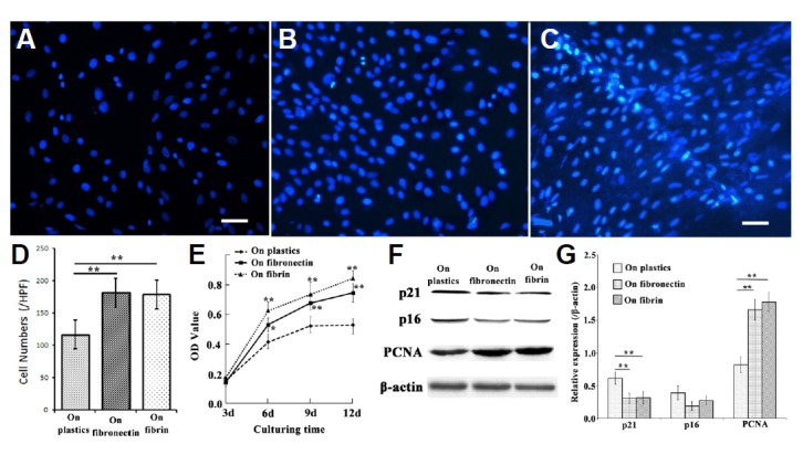
Fig. 4.

Proliferation of EMSCs grown on a plastic surface, fibronectin or the fibrin matrix. Nuclei of EMSCs grown on a plastic surface (A), fibronectin (B), or the fibrin matrix (C). (D) Cell density of EMSCs cultured for 12 days; (E) The results of the MTT assay showed the different rates of proliferation of EMSCs grown on the three substrates. (F) Representative Western blot of cell-cycle markers p21, p16, and PCNA, with β-actin serving as the loading control; (G) quantitative analysis of the Western blot; each bar represents the mean value ± SEM of measurements made in four independent replicates. Bar = 10 μm; *P < 0.05, **P < 0.01.
