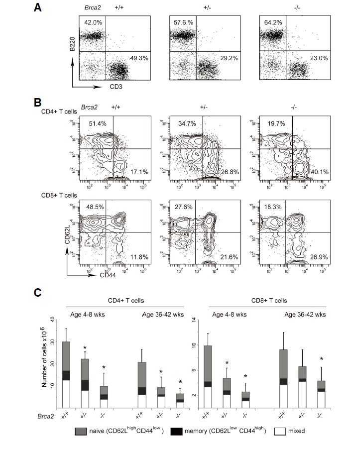
Fig. 3.

Loss of naïve T cells in [Brca2F11/F11; Lck-Cre] mice. Splenic T cells were isolated from 4–8-week-old or 36–42-week-old WT, [Brca2F11/+; Lck-Cre], or [Brca2F11/F11; Lck-Cre] mice. (A) Cells were stained with FITC-ati-B220 and PE-anti-CD3 antibodies. Decreased T cell and increased B cell proportions are shown in the representative plots. Overall number of B cells remained unaltered. (25.9 × 10⁶ ± 8.2; 20 × 106 ± 6.5; 21.7 ± 8 for WT; heterozygous; and homozygous mice respectively) (B) Cells were stained with [PerCP-anti-CD4, APC-anti-CD8, FITC-anti-CD44, and PE-anti-CD62L] antibodies. CD4+ or CD8+ T cells were gated and percentages of cells with CD44low CD62Lhigh expression were calculated from the flow cytometry data. Representative contour plots for activation marker expression (CD44 vs.CD62L) on the gated CD4+ or CD8+ cells are shown. (C) Data were shown as average +/− SD from 3–8 mice for each genotype. P values < 0.05 from t-tests with the WT are marked as asterisks in the figure.
