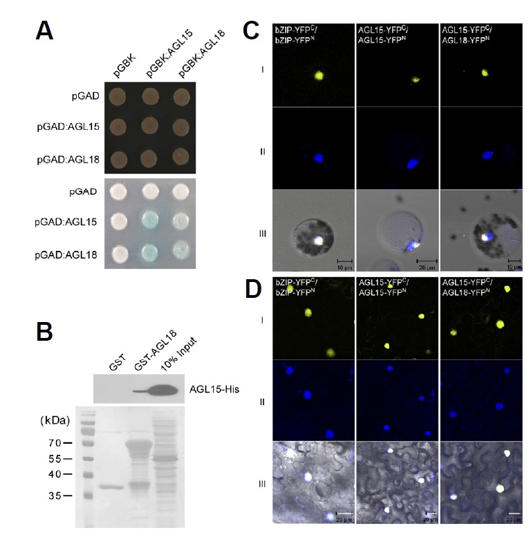
Fig. 5.

Protein–protein interactions of AGL15 and AGL18 in yeast, in vitro, and in vivo. (A) Protein interactions between AGL15 and AGL18 in yeast two-hybrid analysis. Transformed yeast cells were grown on selective SD/-Leu/-Trp (SD-LT) medium (upper panel) and β-galactosidase assay was performed on SD-LT medium (lower panel). (B) Pull-down assay between AGL15-His and GST-AGL18 proteins. The signals were detected using an anti-His antibody. Proteins stained with Ponceau S are shown below. (C and D) Bimolecular fluorescence complementation using AGL18-YFPN and AGL15-YFPC (right column) (YFPN, N-terminal YFP fragment; YFPC, C-terminal YFP fragment) in Arabidopsis mesophyll protoplasts (C), and tobacco leaves (D). Rows I and II indicate YFP fluorescence and the nucleus stained by 4′, 6-Diamidino-2-phenylindole (DAPI), respectively. Row III indicates the merged image of YFP, DAPI signals, and bright field images. bZIP-YFPN and bZIP-YFPC were used for the positive control (Walter et al., 2004).
