
Figure 1.
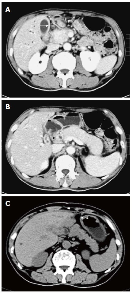
Computed tomography images of autoimmune cholecystocholangitis and pancreatitis. Diffuse gallbladder wall thickening (white arrow) and intrahepatic bile duct dilatation (A), thickening of the common bile duct wall (black arrow) and diffuse swelling pancreas with loss of lobulation (B), and a dramatic recovery in the size of the pancreas after 4 wk of steroid therapy (C).