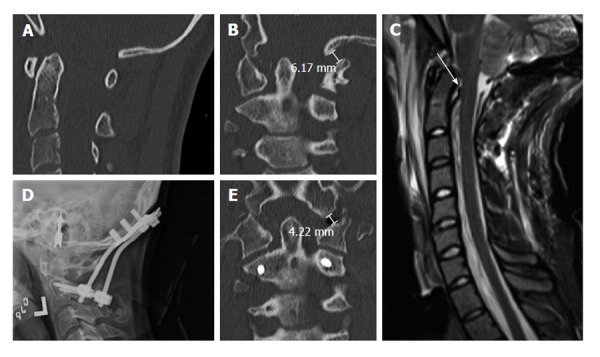
Figure 4.

Nineteen years old woman with traumatic atlanto-occipital dislocation following high-speed motor vehicle accident. A and B: CT of the cervical spine demonstrates no significant abnormalities in the midsagittal plane (A), but clear asymmetry of the occipito-atlantal joints in the coronal plane (B). The left occipital condyle-C1 interval is increased, measuring 6 mm; C: MRI of the cervical spine (T2WI) reveals abnormal signal suggesting disruption of the cruciate ligament; D: Post-operative cervical radiographs show O-C2 fusion using bicortical occipital screws and C2 pedicle screws; E: Post-operative CT of the cervical spine demonstrates reduction of the left occipital condyle-C1 interval to 4 mm. CT: Computed tomography; MRI: Magnetic resonance imaging.
