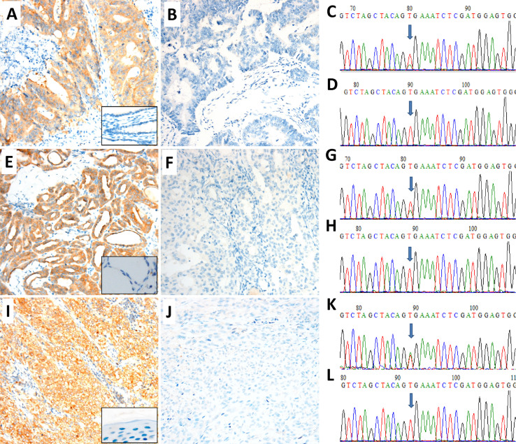
Figure 1. Detection of BRAF mutation in colorectal carcinoma (CRC), papillary thyroid carcinoma (PTC) and melanoma by immunochemistry (IHC) and Sanger sequencing.
Representative images of positive (A, E, I) and negative (B, F, J) for BRAF expression by VE1 IHC. Boxes in A, E, I show the negative controls from their corresponding non-tumor tissues. C, G and K images show a c.1799T > A (p.V600E) point mutation (arrow) of the BRAF gene. D, H and L images show the BRAF mutation (V600E) negative. BRAF Ventana VE1 IHC assay revealed strong expression in BRAF mutation positive patients and no expression in BRAF mutation negative patients in colorectal carcinoma (A–D), papillary thyroid carcinoma (E–H) and melanoma (I–L), respectively. Original magnification ×200.

