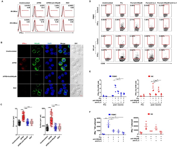
Fig 6. NK cell recall response is influenza-specific and blocked by anti-NKp46.
PBMCs 3 months post-vaccination (subjects #9, #10) were unstimulated or stimulated with A/PR8 (2000 HA/mL) in the presence of 10 μg/mL anti-NKp46, or with RSV (MOI:1) for 18 hours. (A) Histograms depict IFN-γ expression (red) or isotype controls (black) on CD3−CD56+ NK cells by FACS. (B) Representative confocal micrographs of PBMCs (subject #10; 3 months) stained for DAPI (nuclei; blue), NKp46 (green) and IFN-γ (red) expression; colocalization is shown in yellow. Scale bar, 5 μm; original magnification: 63× oil. (C) Diameter and surface area of human NKp46+ cells in (B) (n = 60). **P < 0.01 and ***P < 0.005; one-way ANOVA. (D) PBMCs and purified NK cells at 2 months post-vaccination (subjects #11–13) were restimulated for 18 hours with FluaRIX vaccine (2013–2014) in the presence of 10 μg/mL anti-NKp46 and/or 4 μg/mL neutralizing rat anti—human IL-2 antibodies. Intracellular IFN-γ of CD3−CD56+ human NK cells was detected by FACS. Flu: FluaRIX. (E) Intracellular IFN-γ expression on NK cells detected pre-vaccination (day 0) and at 2 months post-vaccination (subjects #11–13) (PBMCs: blue bars; purified NK cells: red bars). (F) Absolute number of IFN-γ+ cells detected under corresponding conditions at 2 months post-vaccination (PBMCs: blue bars; purified NK cells: red bars) (n = 3). *P < 0.05; Student’s t-test.

