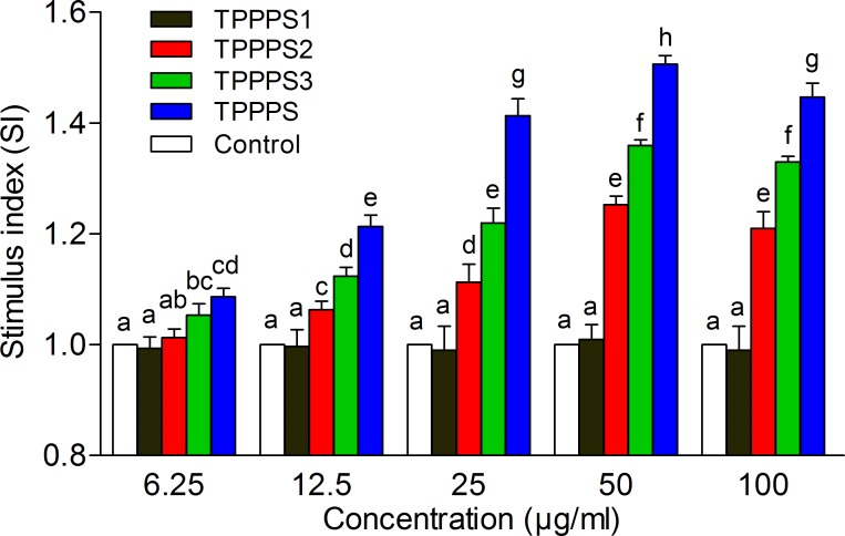
Fig 3. Effects of TPPPS1-3 and TPPPS on splenocyte proliferation in vitro.
Splenocytes were isolated and cultured in 96-well plates. After ConA stimulation, the proliferation was examined by MTT method as described in the Materials and Methods. Stimulus index represents the ratio of absorbance between the experimental group and control group, and the values are presented as mean ± SD from five independent experiments. Different superscripts indicate a significant difference (P < 0.05).

