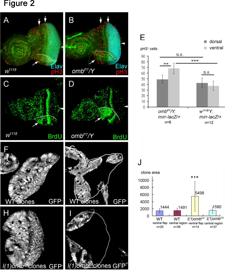
Fig 2. Omb blocks cell proliferation in eye disc.
Cell proliferation was monitored by staining against the mitotic marker phospho-histone 3 (pH3), BrdU incorporation, and by comparing clone size in late third instar eye discs. (A-D) Arrowhead points to the position of the optic stalk. (A) Wild type eye disc showing the two mitotic waves (arrows) labeled by anti-pH3 (red). (B) The omb P7 mutant showed an increased number of pH3-positive nuclei in the ventral eye compared to wild type. (C, D) BrdU incorporation showed an increase of proliferating cells in the ventral flap (arrow) of the omb P7 mutant eye disc (D) compared to the wild type eye disc (C). (E) mirr-lacZ was used to mark the dorsal region. pH3 positive cells were scored in omb P7 /Y; mirr-lacZ/+ and mirr-lacZ dorsal and ventral eyes. In order to include the ventral flap area, the images of several optical sections were merged. The quantification results are summarized in (E). The mitotic cells in ventral eye of omb P7 is significant increased compared to ventral eye in wild type (p<0.05). (F, G) A wild type eye disc with clones (marked by the absence of GFP) at two focal planes to show the central region (F) and the ventral and dorsal flap regions (G). The clones were of similar size in all regions (summarized in J). (H, I) An eye disc with l(1)omb D4 clones (marked by the absence of GFP) at two focal planes to show the central (H) and ventral and dorsal flap regions (I). The wild type clones and l(1)omb D4 mutant clones were induced at the same time. The l(1)omb D4 clones in the ventral flap were on average about 3.5 times larger than omb clones in the central region of the disc or than wild type clones (summarized in J). Differences (*) presented in (E) and (J) are significant (Student′s t-test, *** p<0.001; **, p<0.05).

