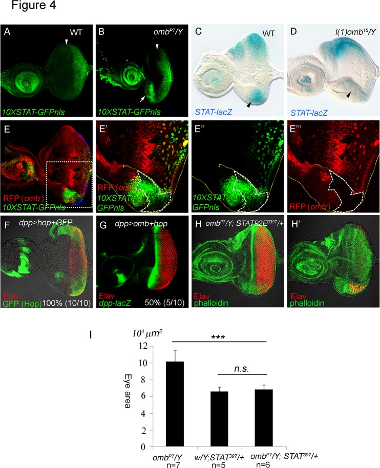
Fig 4. Omb blocks Jak/STAT signaling.
10XSTAT-GFP is a reporter of Jak/STAT signaling [76]. We added a nuclear localizing signal (nls) to obtain 10XSTAT-GFP-nls. (A) 10XSTAT-GFP-nls expression pattern (GFP, green) in wild type third instar eye disc. (B) The 10XSTAT-GFP-nls was ectopically expressed in the ventral eye margin (arrow) in an omb P7 hypomorphic mutant eye disc. (A, B) The position of the MF, based on the DIC image, is marked by an arrowhead. (C) STAT-lacZ is repressed by Jak/STAT signaling. In wild type late third instar eye disc, its expression was strong in the lateral poles and weaker around the DV midline, as reported [57]. (D) In l(1)omb 15 /Y eye discs, STAT-lacZ expression was attenuated in the ventral region. (E-E”’) 10XSTAT-GFP-nls (green) was ectopically induced in l(1)omb D4 mutant clones (clone marked by loss of RFP (red) expression and by dashed line). (E’-E”’) Higher magnification of the square marked in (E). 10XSTAT-GFP-nls was non-autonomously induced by loss of omb in the ventral margin. (F) dpp>hop+GFP caused an enlargement of the eye disc (Elav, red; GFP, green). (G) Coexpression of hop with omb (dpp>omb+hop) could largely rescue the dpp>omb phenotype (dpp-lacZ, green; Elav, red). (H-H’) Reducing STAT dosage in omb P7 /Y; STAT92E 397 /+ larvae reduced the size of the ventral retinal field compared to that in omb P7 /Y (Fig. 2B). Different focal planes of omb P7 /Y; STAT92E 397 /+ were shown in H and H’. The quantified eye areas are summarized in (I).

