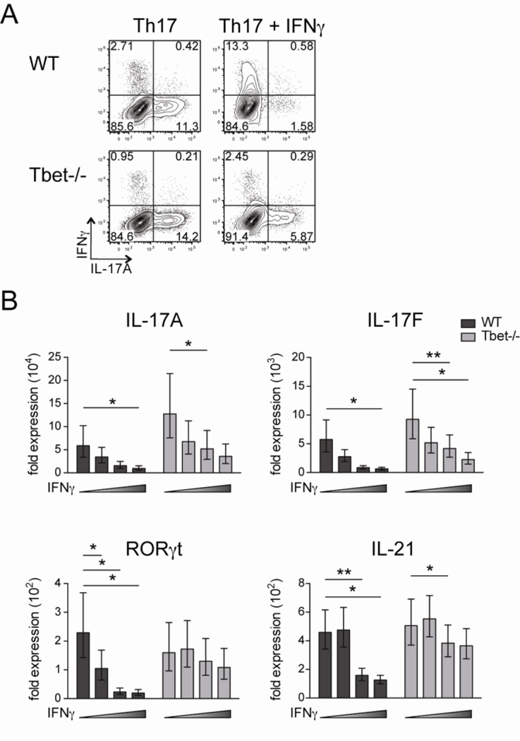
Figure 4. IFNγ suppression of IL-17A/F production independent of Tbet expression.
Naïve CD4 T cells were isolated from WT and Tbet−/− mice, stimulated under Th17 conditions in the presence of irradiated feeder cells for 5 days, and then restimulated with PMA and ionomycin prior to analysis. The cultures contained varying concentrations of IFNγ: 1) 10µg/ml anti-IFNγ mAb, 2) no anti-IFNγ mAb, 3) 100U/ml IFNγ, or 4) 1000U/ml IFNγ. (A) Representative IL-17A and IFNγ staining in CD4 T cells cultured under conditions 1 and 4. Plots are gated on live CD4 T cells. (B) Real-time PCR was performed on live CD4 T cells. Individual gene expression was normalized to β2-microglobulin and shown by the fold expression compared to naive CD4 T cells. Data are combined with three independent experiments and student t test was performed on the value of ΔΔCt.

