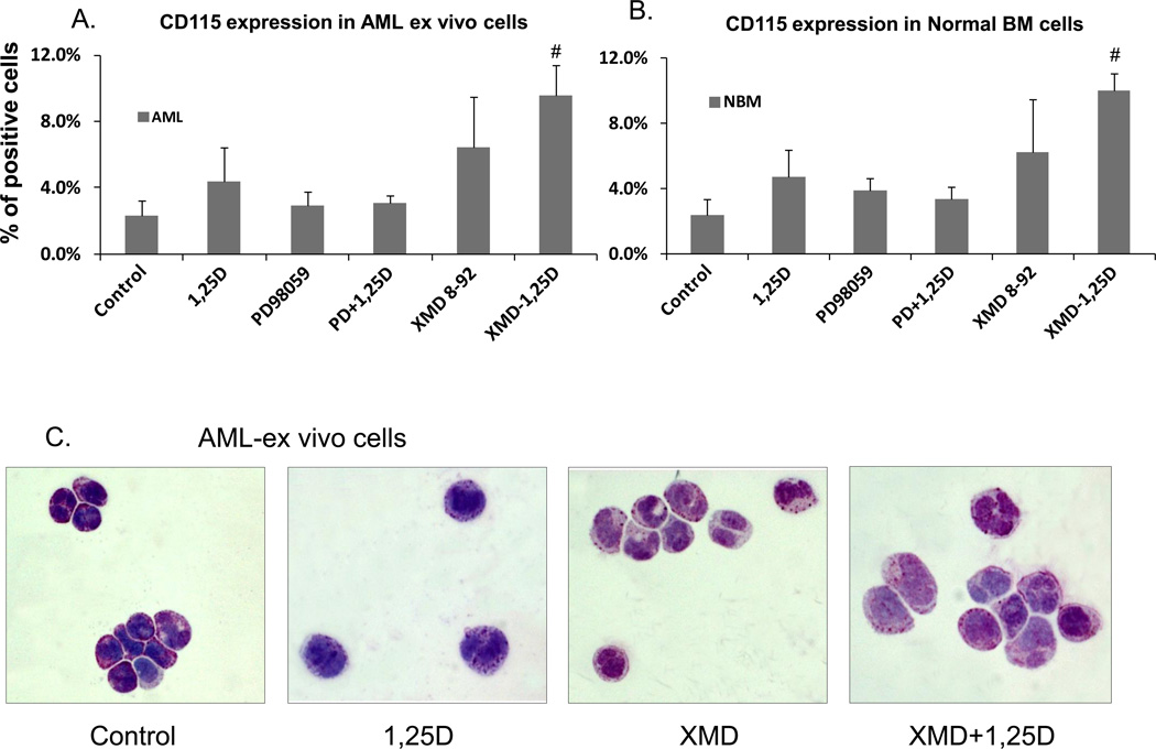
Fig. 6. Macrophage-like phenotype induced by inhibition of ERK5 activity in human AML cells ex vivo.
(A) Flow cytometric determination of surface expression of M-CSFR of AML cells in primary culture. Mononuclear cells were separated from the total cells in specimens of bone marrow, and then subjected to the same treatment as AML cell lines. The expression of M-CSFR (CD115) in primary cultures was determined by flow cytometry. (B) Normal bone marrow cells in primary culture were treated as described for AML cells. # = p<0.05, significantly increased vs. 1,25D-treated group, n=3. (C) Effect of MEK5/ERK5 inhibitors on cell morphology of human AML cells ex vivo. The cells were stained and photographed similarly to those shown in Fig. 2A and B. The cells shown here were from the FAB subtype M2 sample.

