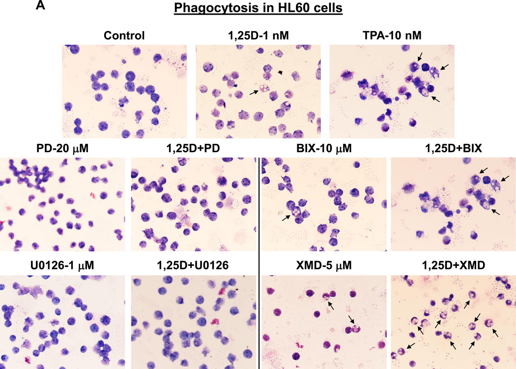
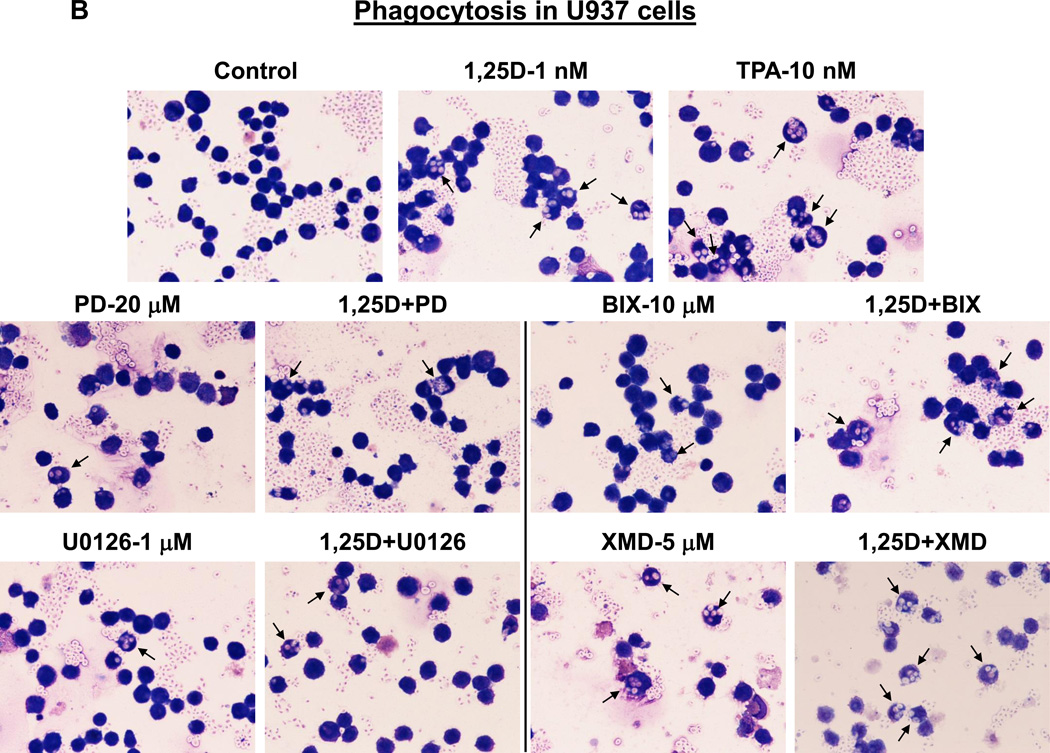
Fig. 7. MEK5/ERK5 inhibitors, but not MEK1/2-ERK1/2 inhibitors, promote phagocytic activity of AML cells.
(A) HL60 cells were pretreated with either MEK5/ERK5 inhibitors, BIX02189 (10 µM) and XMD8-92 (5 µM), or MEK1/2 inhibitors, PD98059 (20 µM) and U0126 (1 µM) for 1 h, then 1,25D (1 nM) was added for an additional 96 h. Cells (5 × 10⁵) were then incubated with opsonized zymosan, smeared on glass slides and stained with Wright-Giemsa. Phagocytosis was determined microscopically at 500× magnification. Arrows indicate examples of cells containing phagocytized zymosan particles. TPA (10 nM) was used here as the positive control for the induction of phagocytosis. (B) U937 cells treated as described above.


