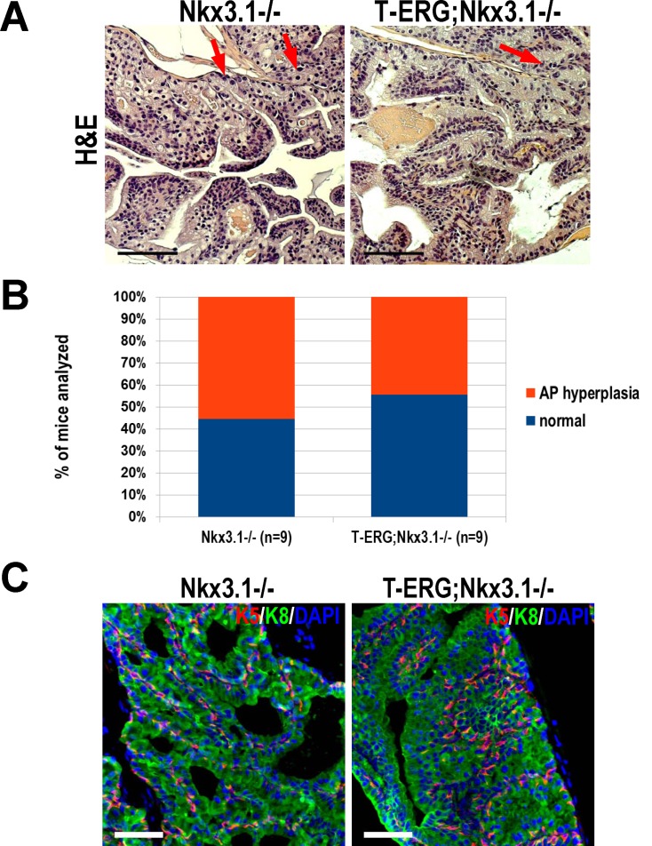
Fig 3. Total Nkx3.1-loss does not cooperate with Tmprss2-ERG gene fusion to promote prostate tumorigenesis.
A. Representative anterior lobe (AP) histology of Nkx3.1 -/- (left) and T-ERG;Nkx3.1 -/- (right) mouse prostates stained with H&E. Scarce pleomorphic nuclei are evident (red arrows). Scale bars represent 100 μm. B. Graphical summary of histological findings of Nkx3.1 -/- and T-ERG;Nkx3.1 -/- male mice. There was no significant difference in AP hyperplasia frequency (p = 0.63). Histology was diagnosed by a trained rodent pathologist. C. IF staining for respective basal keratin 5 (K5, red) and luminal keratin 8 (K8, green) to visualize AP architecture in Nkx3.1 -/- and T-ERG;Nkx3.1 -/- mice. Nuclei counterstained with DAPI (blue). Scale bars represent 50 μm.

