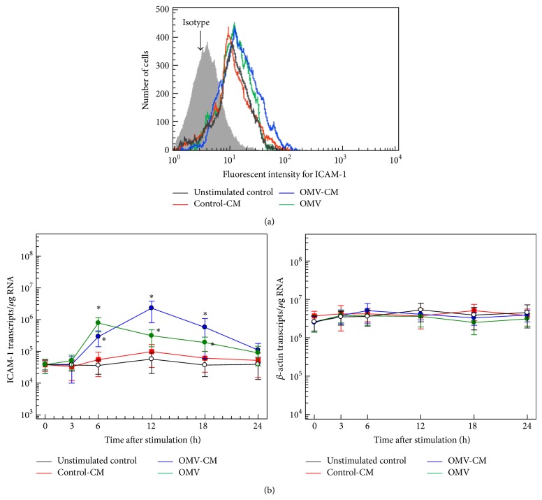
Figure 4.
ICAM-1 expression in gastric epithelial cells stimulated with H. pylori OMVs or OMV-induced culture supernatant. (a) OMV-CM and control-CM were prepared as described in Materials and Methods. Primary human gastric epithelial cells were stimulated with OMVs (200 μg/mL), OMV-CM (50% v/v), or control CM (50% v/v) for 24 h. Cells were stained with a mAb against ICAM-1 and then analyzed using flow cytometry. Results are representative of more than five independent experiments. (b) Time course of ICAM-1 mRNA expression in gastric epithelial cells after stimulation with OMVs, OMV-CM, or control-CM. Cells were stimulated with OMVs (200 μg/mL), OMV-CM (50% v/v), or control-CM (50% v/v) for the indicated periods of time. The expression levels of ICAM-1 and β-actin mRNA were analyzed by quantitative RT-PCR using standard RNAs. Values are expressed as mean ± SD (n = 5). Asterisks indicate statistical significance after comparison with unstimulated controls (P < 0.05).

