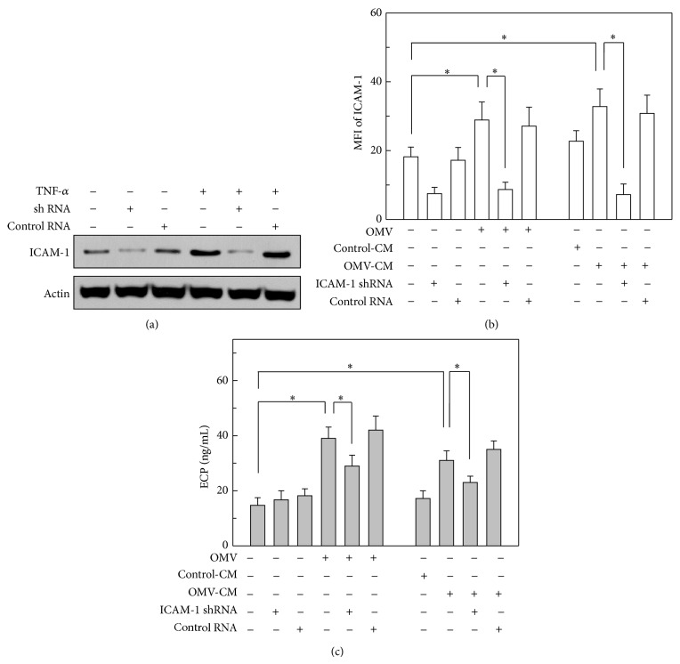
Figure 6.
Relationship between ICAM-1 suppression and ECP release from eosinophils. (a) Primary human gastric epithelial cells were transduced with lentivirus harboring either shRNA directed against ICAM-1 or control shRNA. Transduced cells were then stimulated with TNF-α (20 ng/mL) for 1 h. The cellular levels of ICAM-1 and actin were determined by immunoblot analysis. Results are representative of more than three independent experiments. (b) Transduced or nontransduced gastric epithelial cells were stimulated with OMVs (200 μg/mL), OMV-CM (50% v/v), or control-CM (50% v/v) for 24 h. Cells were stained with a mAb against ICAM-1 and then analyzed by flow cytometry. Data are represented as MFI ± SEM (n = 5). (c) Transduced or nontransduced gastric epithelial cells were exposed to OMVs (200 μg/mL) for 24 h and then washed twice in PBS. OMV-exposed gastric epithelial cells were cocultured with human eosinophils for 24 h (left panel). Transduced or nontransduced gastric epithelial cells were stimulated with control-CM (50% v/v) or OMV-CM (50% v/v) for 24 h and then washed twice in PBS. Finally, human eosinophils were cocultured with either control-CM-stimulated or OMV-CM-stimulated epithelial cells for 24 h (right panel). The concentration of ECP in each culture supernatant was measured by ELISA (mean ± SEM, n = 5). * P < 0.05.

