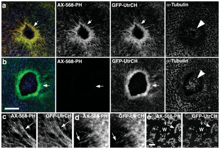
Fig. 1.
Comparison of GFP-UtrCH and fluorescent phalloidin in wounded, fixed oocytes. (a) Low magnification images showing that in samples fixed with an aldehyde-based fixative, fluorescent phalloidin (AX-568-PH) and GFP-UtrCH colocalize, and reveal a striking concentration of F-actin around wounds (arrow). Immunolocalization of tubulin reveals modest preservation of microtubules in the interior of the wound (arrowhead). (b) In samples extracted with methanol after aldehyde fixation, phalloidin staining is completely eliminated, while staining with GFP-UtrCH is preserved. Microtubule preservation is superior under these conditions. (c) High magnification view showing colocalization of fluorescent phalloidin and GFP-UtrCH on cables of F-actin extending away from the wound. (d) High magnification view showing colocalization of fluorescent phalloidin and GFP-UtrCH on actin “fingers” that extend into the wound. (e) Images showing that in cells treated with latrunculin to disrupt F-actin, phalloidin and GFP-UtrCH colocalize in large, unorganized patches around wounds (W). Scale bars is 20 μm.

