Fig. 3.
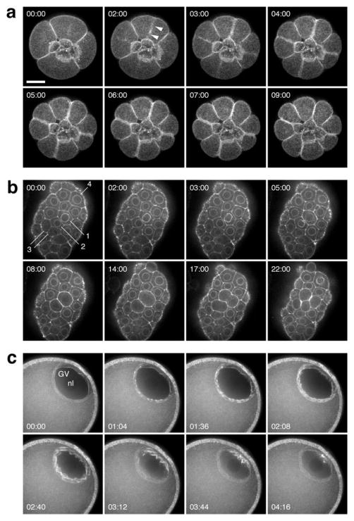
GFP-UtrCH labels F-actin structures in living echinoderm embryos and oocytes. (a) Frames from a 4D sequence of a 16-cell purple urchin embryo expressing GFP-UtrCH from injected mRNA; vegetal view. The four small cells in the center are the micromeres; their sisters, the macromeres, divide in this sequence. Each image is a brightest-point projection of ten 1-μm sections. Probe accumulation (arrowhead) in the equatorial cortex is approximately coincident with the onset of furrowing. (b) Frames from a time-lapse sequence at a single focal plane through the blastula epithelium in a sand dollar embryo. GFP-UtrCH accumulates on the nuclear membrane in inter-phase, brightening just before nuclear envelope breakdown (see cells labeled 1–4; the pair of cells labeled “3” has just divided at the beginning of the sequence, and by 3 min. have accumulated F-actin on the nuclear membrane). (c) Germinal vesicle (GV) breakdown in a sea star oocyte. Actin assembly proceeds in a wave starting at the interior side of the GV. Each timepoint is a projection of eight 1-μm sections. Scale bar is 25 μm.

