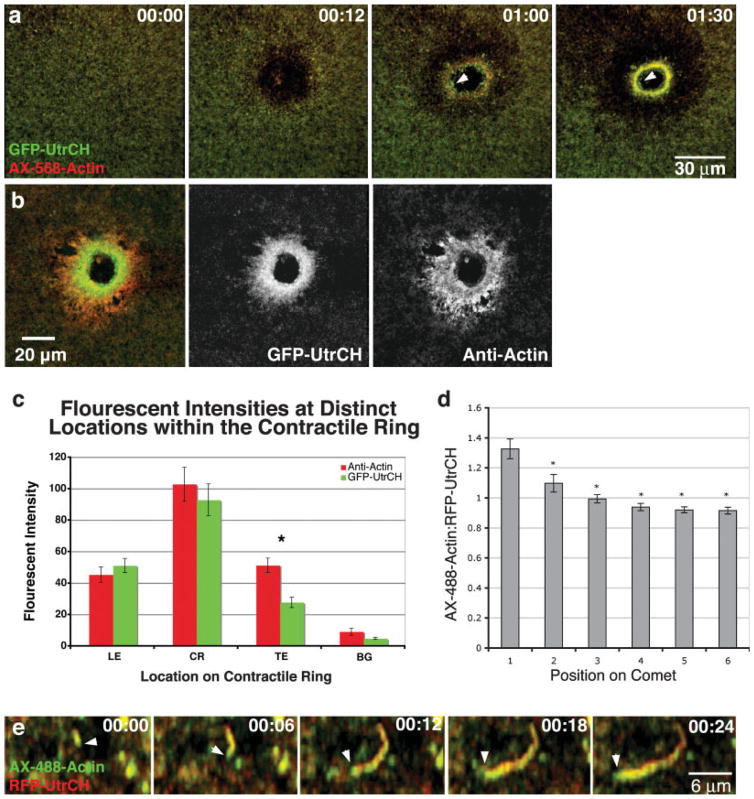
Fig. 4.
Labeling and distinguishing between relatively more dynamic and stable actin structures in Xenopus oocytes. (a) Frames from a 4D movie show that GFP-UtrCH more effectively labels the stable actin that resides on the interior of contractile ring (arrowhead) than AX-568-actin. (b) Comparisons of GFP-UtrCH to total actin distributions by antibody staining in fixed oocytes show few differences at the leading or on the contractile ring but do exhibit differences at the trailing edge. (c) Quantifications of fluorescent intensities at the leading edge (LE), on the contractile ring (CR), and locations away from the contractile structure (BG) showed no differences (P > 0.05, n = 24) between antibody staining and the UtrCH probe. Significant differences in fluorescent intensities were observed at the trailing edge (TE, P < 0.05, n = 24). (d) Quantifications of actin comets from oocytes injected with AX-488 actin and RFP-UtrCH revealed that the ratio of AX-488 actin to RFP-UtrCH is greater at the newly assembled head of the comet than the older tail regions (Asterisks indicate P < 0.05, n = 24). (e) High magnification images showing the growing actin comet head (arrowhead) is labelled more intensely with AX-488 actin than RFP-UtrCH.

