Figure 1.

Ultrastructure and surface morphology of DRibbles.
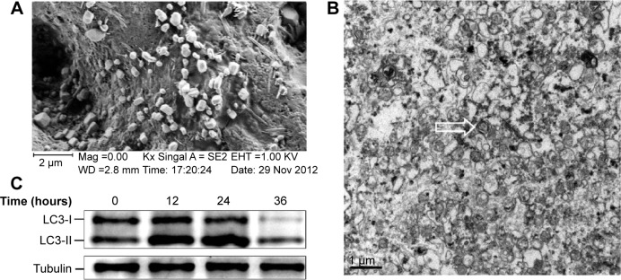
Notes: DRibbles were induced from the SCC7 cell line by rapamycin (100 nM), bortezomib (100 nM), and ammonium chloride (10 mM) for 24 hours. (A) A scanning electron micrograph of DRibbles harvested from SCC7 cells. The diameters of these particles with a relatively smooth surface and spherical structure were in the ranges of 200–500 nm. (B) A transmission electron micrograph of DRibbles. Numerous vesicles with double-membrane structure huddled together, and their dimensions also ranged between 200 nm and 500 nm. The arrow shows autophagosome with the typical double-membrane structure containing undegraded cellular materials. (C) Autophagosomal marker LC3 detected by Western blot analysis. SCC7 cells were treated with 100 nM rapamycin, 100 nM bortezomib, and 10 mM ammonium chloride in complete medium for 12, 24, and 36 hours, respectively. Meanwhile, the untreated SCC7 cells served as control. Cell lysates were prepared from each group. Total proteins were loaded on 4%–12% SDS-PAGE gels and stained with rabbit anti-LC3 antibody for Western blot analysis. LC3-I to LC3-II conversion of SCC7 cells markedly increased after the treatment of rapamycin, bortezomib, and ammonium chloride in a time-dependent manner.
Abbreviations: DRibbles, tumor-derived autophagosomes; SDS-PAGE, sodium dodecyl sulfate polyacrylamide gel electrophoresis.
