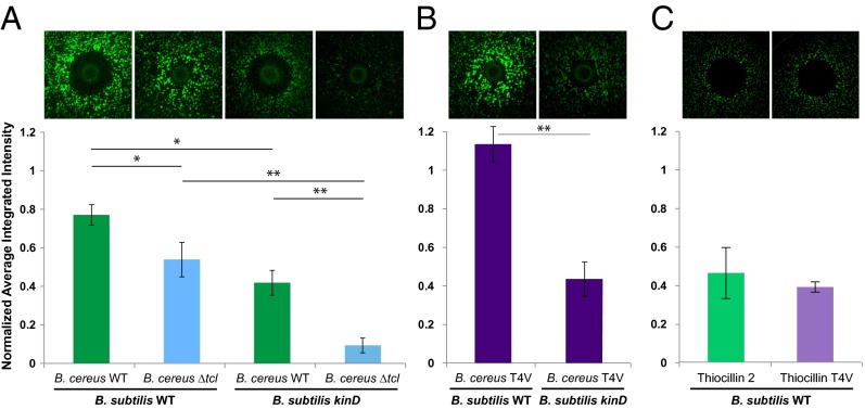
Fig. 2.
Thiocillin contributes to the ability of B. cereus to induce PtapA–yfp gene expression in B. subtilis in a kinD- and antibiotic-independent manner. (A) Colonies of WT and ΔtclE-H B. cereus spotted onto a lawn of WT or kinD B. subtilis PtapA–yfp microcolonies and quantification of the fluorescence (n = 3). (B) Colonies of the thiocillin T4V mutant of B. cereus spotted onto the same B. subtilis lawns, as well as quantification of the fluorescence (n = 3). (C) A total of 450 ng of purified thiocillin or T4V thiocillin spotted onto a lawn of WT B. subtilis PtapA–yfp microcolonies and quantification of the fluorescence (n = 3). The halos visible in C are not due to B. subtilis cells dying, but by being physically moved during spotting of the compound. *P < 0.1; **P < 0.05.

