Fig. 1.
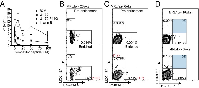
U1-70:I-Ek and P140:I-Ek tetramers specifically detect and enrich MRL/lpr CD4+ T cells. (A) Competition assay for peptide binding to I-Ek. CH27 B cells were pulsed with a suboptimal concentration of MCC and increasing amounts of competitor peptide. IL-2 production from 2B4 T cells, cultured with peptide-pulsed CH27 B cells, was measured by ELISA. Data are representative of two individual experiments. Error bars are derived from triplicate conditions. (B and C) Tetramer staining of CD4+ T cells from MRL/lpr lymph nodes with U1-70:I-Ek (B) and P140:I-Ek (C) before enrichment (Upper) and after positive selection enrichment of tetramer-positive cells using anti-PE or anti-APC magnetic microbeads (Lower). Values in black are percentages in the gate shown; values in red are the calculated numbers of tetramer-positive cells among 106 CD4+ T cells. (D) U1-70:I-Ek and P140:I-Ek tetramers did not costain CD4+ T cells from MRL/lpr mice at 18 or 6 wk of age. Flow cytometry plots show the enrichment of CD4+ T cells from two individual MRL/lpr mice that were processed, stained with tetramers, and enriched within the same experiment. Results are representative of 10 mice tested. Cells are gated on MCC:I-Ek-negative CD4+ T cells.

