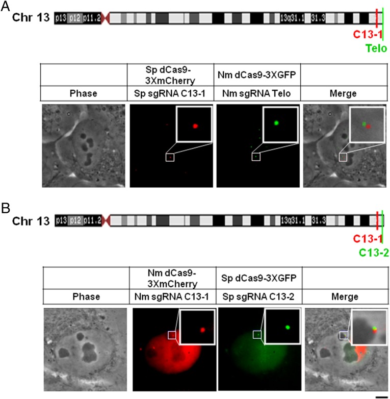
Fig. 4.
Spatial resolution of subtelomeric loci in chromosome 13 and the adjacent telomere. (A) Diagram of the locations of C13-1 and the telomere on the long arm of chromosome 13. Sp dCas9-3xCherry (Middle Left) and Nm dCas9-3xGFP (Middle Right) were coexpressed in U2OS cells together with their cognate sgRNAs (Sp sgRNA-C13-1 and Nm sgRNA-Telo). Shown are a phase-contrast image (Left), the Cherry and GFP fluorescence images (Middle), and the merged image (Right). (B) Diagram of the locations of C13-1 and C13-2. Nm dCas9-3xCherry (Middle Left) and Sp dCas9-3xGFP (Middle Right) were coexpressed in U2OS cells together with their cognate sgRNAs (Nm sgRNA-C13-1 and Sp sgRNA-C13-2). Shown are a phase-contrast image (Left), the Cherry and GFP fluorescence images (Middle), and the merged fluorescence image (Right). In the right panel, most of the uniformly dispersed fluorescence background (Middle Left) was removed by increasing the threshold to facilitate observation of the merged signal. (Scale bar: 5 μm.)

