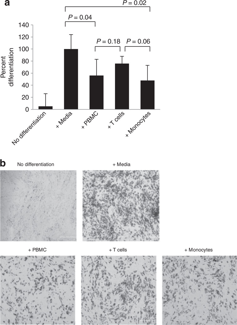
Figure 2.
Differentiation of human subcutaneous preadipocytes for 2 weeks following 6 days of coculture with PBMCs, T cells, or monocytes (i.e., after 6 days of coculture, PBMCs, T cells, or monocytes were removed and preadipocytes were differentiated in the absence of these cells). (a) Quantification of preadipocyte differentiation after 2 weeks following hormonal induction. Adipocyte lipids were stained with Oil Red O and four random fields of observation from each condition were recorded. Pixel values of Oil Red O-stained areas were calculated with Adobe Photoshop software and averaged. Normal differentiation with control media was established as 100%. N = 3, error bars are means ± s.d. (b) Representative phase contrast microscopy images (×40) of Oil Red O-stained preadipocytes after 2 weeks of differentiation. PBMC, peripheral blood mononuclear cell.

