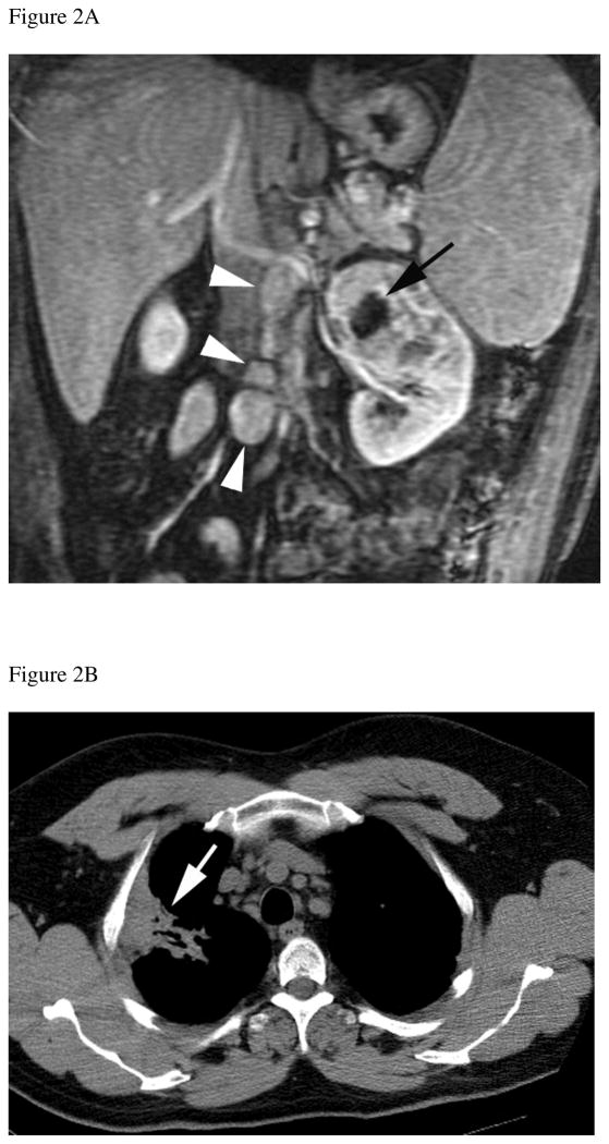
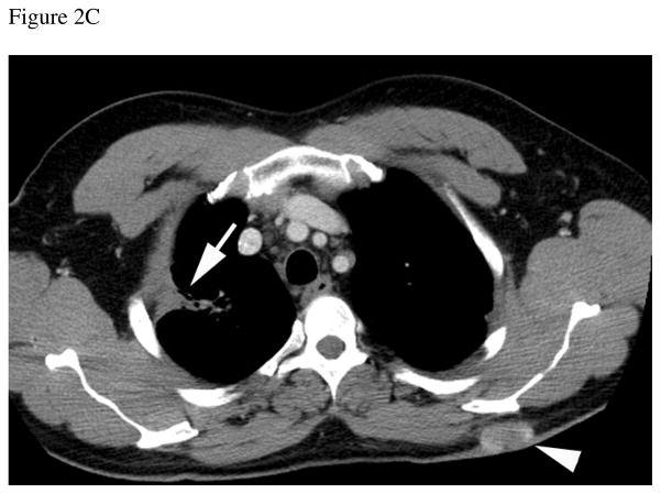
Figure 2.
Thirty-eight year-old male with history of sarcoidosis and left renal mass. A) Gadolinium-enhanced coronal T1-weighted gradient echo MR image shows a 6.5 cm mass in the upper pole of the left kidney. A central non-enhancing area of necrosis is present (black arrow). Multiple enlarged retroperitoneal lymph nodes (arrowheads) are also seen. After nephrectomy, histopathology analysis revealed clear cell carcinoma, Fuhrman grade II. Analysis of the retroperitoneal lymph nodes demonstrated numerous non-necrotizing granulomas but no evidence of metastatic disease. B) Axial CT image of the chest obtained 1 year after nephrectomy shows stable lung parenchymal consolidation in the right upper pole (arrow) compared to prior CT examinations (not shown) consistent with sarcoidosis. C) Axial contrast-enhanced CT at the same level as B, obtained 3 years after nephrectomy, shows persistent lung consolidation (arrow) and a new enhancing soft tissue nodule in the subcutaneous fat of the back (arrowhead). Histopathologic evaluation of this soft-tissue nodule after surgical excision confirmed metastatic clear cell renal cell carcinoma


