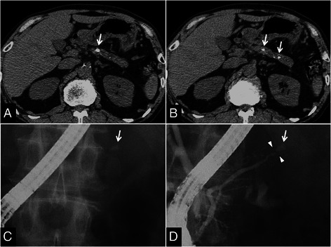
Figure 1.

CT and ERCP findings in a 66-year-old man whose pancreatic stone was treated with ESWL to preserve pancreatic function. (A), (B) CT before ESWL showing the pancreatic stone and pancreatic atrophy (arrows). (C), (D) ERCP before ESWL identifying the obstructing X-ray-positive stone in the MPD (arrows) and pancreatic duct stenosis proximal to the pancreatic calculus (arrowheads). Pre-pancreatograpy (C) and post-pancreatography (D) images.
