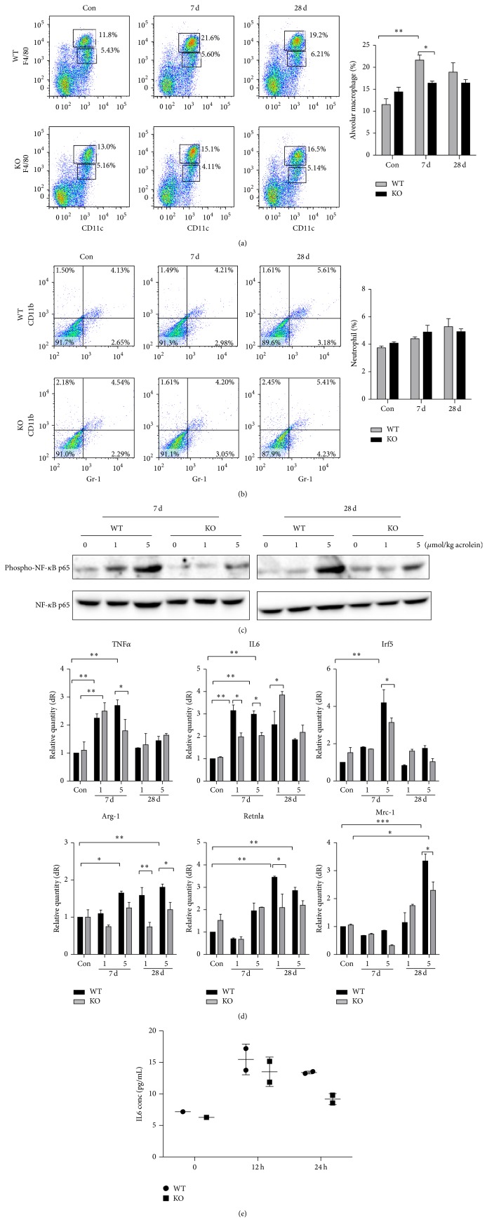
Figure 4.
Low level of pulmonary inflammation in GADD34-knockout mice induced by acrolein. Lungs were collected from wild-type and GADD34-knockout mice at days 7 and 28 after 5 μmol/kg acrolein instillation. (a) Alveolar macrophages as F4/80highCD11c+ and (b) neutrophils as Gr-1+CD11b+ were confirmed by FACS. (c) The expression of phospho-NF-κB p65. (d) The expressions of macrophage type I markers, TNFα, IL-6, and Irf5, and macrophage type II markers, Arg-1, Mrc-2, and Retnla, were analyzed by quantitative real-time PCR. (e) Wild-type and GADD34-knockout mice macrophages were cultured in 12-well plastic plates and stimulated with 10 μM acrolein for 12 and 24 h. Supernatants were taken and IL-6 expression was analyzed by ELISA. Data shown are the mean ratios ± SE of three separate experiments. Data are represented as means ± s.e.m. * P < 0.05, ** P < 0.01.

