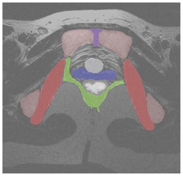
Figure 3.
The T2-weighted axial MRI slice from Figure 1, shaded with the consensus segmentation of each organ as computed by STAPLE. Legend: red, obturator internus; green, levator ani; blue, vagina; white, rectum; gray, bladder neck; violet, symphysis; pink, pelvic bones.

