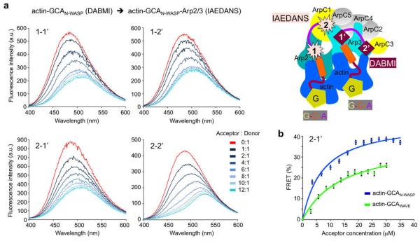
Figure 6.
teady state FRET analysis of actin-GCA binding to Arp2/3 complex. (a) Fluorescence quenching during titration of acceptor-labeled actin-GCAN-WASP into donor-labeled 1:1:1 actin-GCAN-WASP-Arp2/3 complex. Probe sites and titration ratios are indicated. A cartoon representation illustrates the relative positions of the donor (IAEDANS) and acceptor (DABMI) probes in these experiments. (b) FRET as a function of acceptor concentration for the 2-1’ titrations (with smallest red shift) of actin-GCAN-WASP (blue) and actin-GCAWAVE (green, experiment shown in Supplementary Fig. S7). Data points represent mean ± s.e.m. Data fitting resulted in Kd values of 7 and 14 μM for actin-GCAN-WASP and actin-GCAWAVE, respectively.

