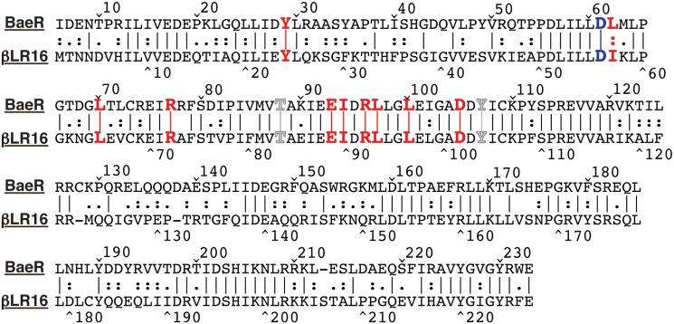
Fig 2. Amino acid alignment of the BaeR response regulator of E. coli and the response regulator from βLR16.
Alignment was performed with MegAlign from the Lasergene software package from DNASTAR (Madison, WI) using the Lipman-Pearson alignment tool (Ktuple:2; Gap Penalty: 4; Gap Length Penalty: 12). Levels of similarity between the sequences are indicated as lines (identical) or dots (similar). The conserved aspartate residue that is phosphorylated is indicated in blue (Asp 56 in βLR16). Switch residues are indicated in gray (Thr 83 and Tyr 102 in βLR16), and amino acids from BaeR involved in domain swap interactions, and their βLR16 counterparts, are shown in red.

