
Figure 4.
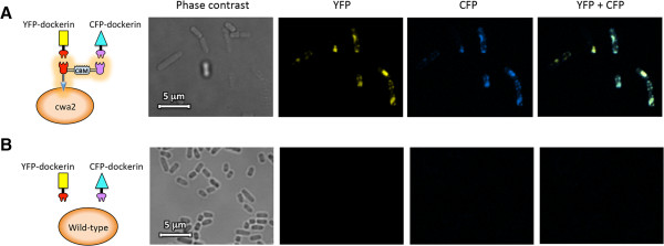
Confocal fluorescence microscopy images of Scaf · AT in cwa2-carrying strain (A) or wild-type cells (B) after binding interaction with mVenus yellow fluorescent protein (YFP) and mCerulean cyan fluorescent protein (CFP) fluorophores fused to respective dockerin modules. Bar, 5 μm.