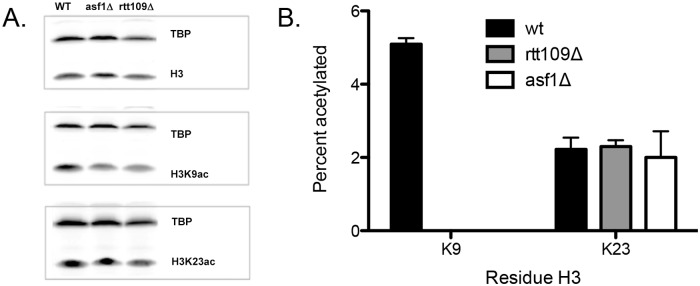
Fig 6. Changes in vivo due to the loss of either Rtt109 or Asf1.

(A) Western blots using antibodies for H3, H3K9 or H3K23, using whole cell extracts from wild type (wt), rtt109Δ, and asf1Δ strains. (B) Determination of the percentage of acetylation at position H3K9 or H3K23 in wt, rtt109Δ, and asf1Δ strains using the targeted MS-based method. The error bar represents the standard error in acetylation percentage.
