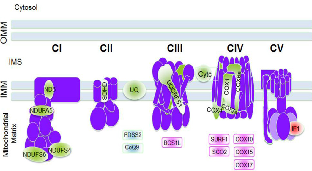
Figure 1. Mouse models of mitochondrial diseases.
Mouse models with defects in OXPHOS complexes subunits, electron carriers and assembly factors are summarized in the figure. In light green are represented the mouse models related to subunits of the electron transport chain (ND6, NDUFA5, NDUFS4, NDUFS6, SDHD, UQCRFS1, COX1, COX4, COX6a and COX7a), the electron carriers ubiquinone (UQ) or CoQ and cytochrome c (Cytc). In light blue are the mouse models of enzymes of the CoQ biosynthetic pathway (PDSS2 and CoQ9). In light pink are represented those mouse models of assembly factors for CIII and CIV (BCS1L, SURF1,SCO2, COX10, COX15, and COX17) and in red the mouse models of IF1, the natural inhibitor of CV.
OMM: outer mitochondrial membrane, IMS: inter membrane space; IMM: inner mitochondrial membrane

