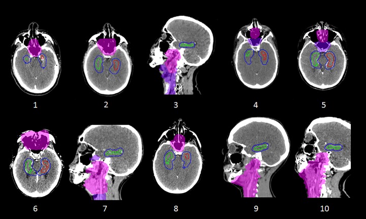
Fig 2. Disposition of PTVs and hippocampal PRVs for the 10 cases.
Case numbering is as per Table 1. PTV1 is shown as a light pink colourwash, PTV2 as a purple colourwash, while the left hippocampus, right hippocampus and hippocampal PRVs are illustrated as red, green, and blue contours respectively. Axial or sagittal views are shown for each case according to the plane that transects both volumes.

