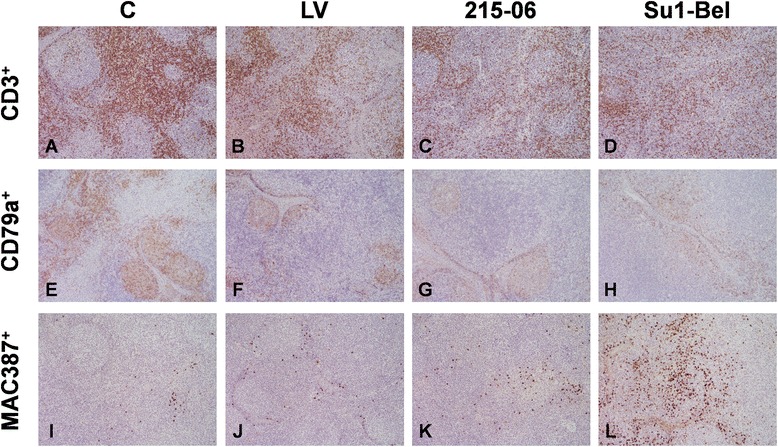
Figure 3.

Representative images of CD3, CD79a and MAC387 IHC staining in mediastinal lymph node. CD3 (A, B, C, D), CD79a (E, F, G, H) and MAC387 (I, J, K, L) in mediastinal lymph nodes of control pigs (A, E, I) and infected with LV (B, F, J), 215–06 (C, G, K) and SU1-Bel (D, H, J) strains, at 7 dpi. An increased in the number of CD3+ cells is observed in the lymphoid follicles from all infected groups together with a depletion of these cells in the interfollicular areas. A decrease in the number of CD79a cells in the follicles is also observed in all the infected groups. A substantial increase in the number of MAC387 is observed in the follicles and interfollicular areas from SU1-Bel infected animals (L) together with a mild increase in the LV (J) and 215–06 (K) groups. Original magnification: 20x.
