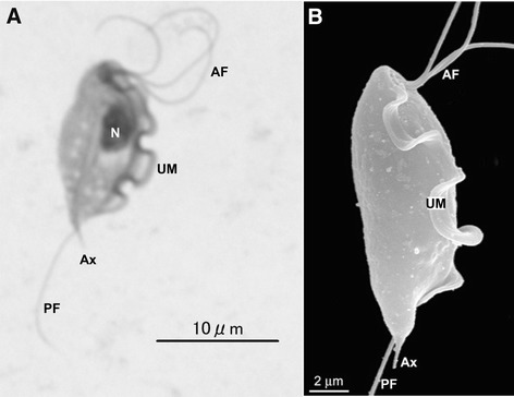
Figure 1.

Tritrichomonas foetus trophozoites. A: Giemsa-stain. B: scanning electron microscopy. AF: anterior flagella; Ax: axostyle; N: nucleus; PF: posterior flagellum; UM: undulating membrane. A and B: reproduced from Figure 1 of Doi et al. ([80]) and Figure 1b of Midlej et al. [81], respectively, with permission.
