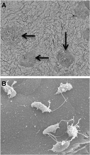
Figure 3.

Scanning electron microscopy of Tritrichomonas foetus adhesion to porcine intestinal epithelial cell (IPEC)-J2 monolayers. A. Aggregates of trophozoites adhering to IPEC-J2 monolayers (arrows). B. Six trophoziotes adhering to a single IPEC-J2 cell. Reproduced with permission [47].
